 |
Case Report
Volar dislocation of the second and third carpometacarpal joints: A case report and detailed mechanism of injury
1 MS, Medical Student, Florida State University College of Medicine, Tallahassee, FL, USA
2 MS, Medical Student, University of Puerto Rico School of Medicine, San Juan, PR, USA
3 MD, Orthopaedic Hand Surgeon, Florida Orthopaedic Institute, Tampa, FL, USA
Address correspondence to:
Nicholas D Thomas
MS, 1115 West Call Street, Tallahassee, FL,
USA
Message to Corresponding Author
Article ID: 100028Z14NT2022
Access full text article on other devices

Access PDF of article on other devices


How to cite this article
Thomas ND, Rodríguez SF, Lopez P. Volar dislocation of the second and third carpometacarpal joints: A case report and detailed mechanism of injury. J Case Rep Images Orthop Rheum 2022;6:100028Z14NT2022.ABSTRACT
Introduction: Carpometacarpal (CMC) joint dislocations are rare injuries that are difficult to diagnose radiographically. The third metacarpal articulates with the capitate bone at the wrist more proximally, and forms the strongest of all CMC joints. Therefore, dislocation of this joint may lead to an increased risk of instability and failed reduction. The present case highlights a unique injury pattern that is frequently overlooked.
Case Report: A 28-year-old man presented for examination of his left hand after a high velocity fall while skateboarding. A video recording provided at the office visit depicted the inciting injury. The converging forces during the fall caused hyperextension of the wrist at the base of the second and third metacarpal bones. Initially after this incident he arrived at the emergency department where computed tomography (CT) scans were interpreted as normal. Two weeks later, he presented in clinic with increased weakness and pain and X-rays showed dislocation of the second and third CMC joint. The following day, open reduction and external fixation (ORIF) was performed. Eight weeks after surgery, the patient presented with excellent range of motion, and X-rays of the hand showed stable articulation of the second and third CMC joints.
Conclusion: Few cases of volar dislocations of the second and third CMC joints have been reported. Treatment for CMC joint dislocation is controversial. However, early ORIF has shown excellent clinical outcomes for patients with multiple CMC dislocations. Thorough clinical and radiographic examination is necessary to prevent chronic dislocation which may lead to diminished grip strength, nerve damage, and carpal instability.
Keywords: Carpometacarpal, CMC, Dislocation, Volar, Wrist
Introduction
Carpometacarpal (CMC) joint dislocations are rare injuries that are difficult to diagnose radiographically [1]. These injuries are frequently missed or delayed in diagnosis due to concomitant hand swelling and superimposed hand bones [2],[3]. Most dislocations occur at the fourth and fifth digits, with the fifth digit being reportedly involved in up to 80% of cases [4],[5],[6]. Dislocation of the second and third CMC joints is even less common. The third metacarpal articulates with the capitate bone at the wrist more proximally, and forms the strongest of all CMC joints [3]. Therefore, dislocation of this joint may lead to increased risk of instability and failed reduction [7],[8].
The second and third CMC joints at the index and long finger are essentially immobile, allowing only about 1–3 degrees of motion [4],[9]. Volar and dorsal ligaments, as well as the surrounding hand and wrist muscles provide dynamic stabilization at this joint [10]. Both dorsal and volar dislocations have been reported in the literature based on direction of force. Previous reports suggest that dorsal CMC joint dislocations occur at higher rates [3],[10]. Few cases of volar dislocations of the second and third CMC joints have been reported, with most cases occurring via high-energy trauma during motor vehicle accidents [11],[12],[13],[14]. However, the present case highlights a unique and detailed mechanism contributing to a rare injury pattern that is frequently overlooked.
Case Report
A 28-year-old man presented for examination of his left hand after a high velocity fall while skateboarding. A video recording provided at the office visit depicted the patient riding his skateboard over the length of a grind rail, in the “regular” skateboard stance, with his left foot forward (Video 1). His skateboard suddenly slipped from underneath him during his stunt and he began falling towards the rail. At that point, he reached down between his legs to grab the rail with his arm fully extended using his left hand to brace for impact. As he grabbed the rail with his left hand, he increased axial compression onto the wrist extended at 90° while the weight of his body moved inward and inferiorly onto his wrist below him. His left leg also came forward and forcefully pushed on his arm just below the posterior aspect of the elbow. The converging forces applied to the wrist in this position caused an abrupt dorsiflexion of the hand leading to hyperextension of the wrist with slight radial deviation. The direct, high velocity impact at the CMC joint in the present case caused the base of the second and third metacarpal bones to shift volarly.
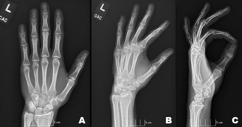
Initially after his incident he arrived at the emergency department where CT scans were taken of the left upper extremity, and interpreted as no acute fracture or subluxation. Two weeks after the inciting event, he presented in our clinic with increased weakness and pain. On physical exam, the patient had diffuse swelling of the left hand and wrist with tenderness over the second and third CMC joints. After clinical examination the patient underwent radiographic evaluation. Anterior posterior (AP), lateral, and oblique views of the wrist and hand were taken and reviewed by a board-certified orthopedic hand surgeon. Lateral view indicated a dislocation of the second and third CMC joints. Further radiographic evaluation showed widening of the second and third CMC joint space, but no accompanying fractures or associated injuries of the hand or wrist (Figure 1).
The following day, open reduction and external fixation (ORIF) of the left index and long fingers was performed. The metacarpals were stably reduced, and fixation was maintained by two 0.045″ Kirschner wires (pins) driven through the third metacarpal, across the CMC joint, into the capitate and trapezoid bones, respectively (Figure 2). The patient was placed into a volar wrist splint immediately after surgery and switched into a thermoplastic splint at his first post-operative visit three days later. Active finger movements were started on first postoperative day to prevent stiffness. Sutures were removed after two weeks, and pins were removed at the 4-weeks postoperative visit. At 8-weeks follow-up, the patient presented with excellent strength and range of motion, and X-rays of the hand (3-views) showed stable articulation with no subluxation of the second and third CMC joints (Figure 3).
Access video on other devices


Discussion
Carpometacarpal dislocations have been well characterized in the literature with an incidence of less than 1% of all hand injuries [1],[4],[15]. Previous studies show the fourth and fifth CMC joints are most frequently involved in dislocation injuries due to high mobility and loose ligamentous attachments [4],[5],[6]. However, dislocation of the second and third CMC joints are rarely reported [11]. Nonetheless, the index and long finger metacarpals are important pillars of the hand with increased rigidity [9],[16]. Additional insertion of the flexor carpi radialis and extensor carpi longus and brevis muscles allow minimal range of motion around these joints [14]. When the patient grabbed the rail with the wrist radially deviated, the flexor carpi radialis muscle was activated which attaches volarly at the base of the second and third metacarpals. The contraction of this muscle forcefully pulled the metacarpals, while simultaneous hyperextension at the wrist pushed the metacarpals in the same direction, leading to volar dislocation.
The third CMC joint is historically known for its anatomic positioning more proximal than its co-parts [11]. In cases of multiple CMC joint dislocations, previous studies state the third CMC joint is a “key-stone” and must be re-set before others [3]. The third metacarpal articulates with the capitate bone, which is the center piece of the wrist and largest carpal bone [3]. Notably, all the muscles responsible for movement at the wrist cross axes of rotation located at the capitate bone which may lead to greater instability upon dislocation [14].
Treatment for CMC joint dislocation is controversial [15] and may be treated conservatively via close reduction and splint immobilization, or operatively via ORIF with Kirschner wires, as seen in the present case. However, closed reduction has reportedly higher risk of re-dislocation and non-union of accompanying fractures, if present [1]. Operative management is favored due to opportunity for debridement of the joint space, drainage of local hematoma, and repair of damaged tendons [8]. Early ORIF has shown excellent clinical outcomes for patients with multiple CMC dislocations [1],[15].
The present case is representative of the commonly missed emergency room (ER) diagnosis of a CMC dislocation, usually attributed to posttraumatic edema [3]. Radiographic indications for assessment are consistent with AP, lateral, and oblique views being gold standard for identifying CMC dislocations [8],[12]. Computed tomography (CT) may be used for diagnosis in the case of occult X-ray findings or accompanying carpal bone fractures [4]. However, in this case, CT scans were interpreted as normal. Thorough clinical and radiographic examination is necessary to prevent misdiagnosis leading to chronic dislocation, nerve damage, and carpal instability [9],[15].
Conclusion
We report a rare case of volar CMC dislocations of the index and long finger metacarpals successfully treated with ORIF. This case is of interest as the exact incident was recorded on video, providing a clear understanding of the forces that caused the injury. Due to the rarity of this injury, it is subject to missed diagnosis in the emergency setting. This study highlights a unique mechanism of injury to a key stabilizing joint of the wrist.
REFERENCES
1.
Sharma AK, John JT. Unusual case of carpometacarpal dislocation of all the four fingers of ulnar side of hand. Med J Armed Forces India 2005;61(2):188–9. [CrossRef]
[Pubmed]

2.
Jumeau H, Lechien P, Dupriez F. Conservative treatment of carpometacarpal dislocation of the three last fingers. Case Rep Emerg Med 2016;2016:4962021. [CrossRef]
[Pubmed]
3.
Pundkare GT, Patil AM. Carpometacarpal joint fracture dislocation of second to fifth finger. Clin Orthop Surg 2015;7(4):430–5. [CrossRef]
[Pubmed]
4.
Desai B, Nammour M, Warren M, Sumarriva G, Sisco-Wise L. Isolated volar dislocation of the fifth carpometacarpal joint. Ochsner J 2020;20(2):215–8. [CrossRef]
[Pubmed]
5.
Fischer JW, Waseem M, Gambhir A, Creedon RJ. Ulnopalmar dislocation of the fifth carpometacarpal joint. A rare injury. Acta Orthop Belg 2002;68(2):175–7.
[Pubmed]
6.
Kural C, Başaran SH, Ercin E, Bayrak A, Bilgili MG, Baca E. Fourth and fifth carpometacarpal fracture dislocations. Acta Orthop Traumatol Turc 2014;48(6):55–60. [CrossRef]
[Pubmed]
7.
Halilaj E, Rainbow MJ, Got C, et al. In vivo kinematics of the thumb carpometacarpal joint during three isometric functional tasks. Clin Orthop Relat Res 2014;472(4):1114–22. [CrossRef]
[Pubmed]
8.
Hartwig RH, Louis DS. Multiple carpometacarpal dislocations. A review of four cases. J Bone Joint Surg Am 1979;61(6A):906–8.
[Pubmed]
9.
Kollitz KM, Hammert WC, Vedder NB, Huang JI. Metacarpal fractures: Treatment and complications. Hand N Y 2014;9(1):16–23. [CrossRef]
[Pubmed]
10.
Kumar R, Malhotra R. Divergent fracture-dislocation of the second carpometacarpal joint and the three ulnar carpometacarpal joints. J Hand Surg 2001;26(1):123–9. [CrossRef]
[Pubmed]
11.
Kumar P. Dislocation of second and third carpometacarpal joints along with fracture of first metacarpal—possible mechanisms. J Hand Microsurg 2010;2(2):85-6. [CrossRef]
[Pubmed]
12.
Silk G, Vetharajan N, Nagata H. Volar dislocation of the second and third carpometacarpal joints – The Lisfranc injury of the hand? Hand Surg Rehabil 2018;37(5):320–3. [CrossRef]
[Pubmed]
13.
Weiland AJ, Lister GD, Villarreal-Rios A. Volar fracture dislocations of the second and third carpometacarpal joints associated with acute carpal tunnel syndrome. J Trauma 1976;16(08):672–5. [CrossRef]
[Pubmed]
14.
Zaizi A, El Yaacoubi T, El Bahraouy A, et al. Pure divergent dislocation of the index and middle finger carpometacarpal joints: A rare case. Trauma Case Rep 2019;23:100222. [CrossRef]
[Pubmed]
15.
Bell T, Chinchalkar SJ, Faber K. Postoperative management of carpometacarpal joint fracture dislocation of the hand: A case report. Can J Plast Surg 2010;18(3):e37–40.
[Pubmed]
16.
Taleisnik J. The ligaments of the wrist. J Hand Surg 1976;1(2):110–8. [CrossRef]
[Pubmed]
SUPPORTING INFORMATION
Author Contributions
Nicholas D Thomas - Acquisition of data, Analysis of data, Drafting the work, Revising the work critically for important intellectual content, Final approval of the version to be published, Agree to be accountable for all aspects of the work in ensuring that questions related to the accuracy or integrity of any part of the work are appropriately investigated and resolved.
Sebastián Frontera Rodríguez - Acquisition of data, Analysis of data, Revising the work critically for important intellectual content, Final approval of the version to be published, Agree to be accountable for all aspects of the work in ensuring that questions related to the accuracy or integrity of any part of the work are appropriately investigated and resolved.
Peter Lopez - Conception of the work, Design of the work, Revising the work critically for important intellectual content, Final approval of the version to be published, Agree to be accountable for all aspects of the work in ensuring that questions related to the accuracy or integrity of any part of the work are appropriately investigated and resolved.
Guaranter of SubmissionThe corresponding author is the guarantor of submission.
Source of SupportNone
Consent StatementWritten informed consent was obtained from the patient for publication of this article.
Data AvailabilityAll relevant data are within the paper and its Supporting Information files.
Conflict of InterestAuthors declare no conflict of interest.
Copyright© 2022 Nicholas D Thomas et al. This article is distributed under the terms of Creative Commons Attribution License which permits unrestricted use, distribution and reproduction in any medium provided the original author(s) and original publisher are properly credited. Please see the copyright policy on the journal website for more information.


