
 |
Case Report
Diffuse alveolar hemorrhage secondary to antiphospholipid syndrome presenting as recurrent multifocal pneumonia
1 Baylor Scott and White Medical Center, Round Rock, TX, USA
2 Texas A&M Naresh K. Vashisht College of Medicine, Bryan, TX, USA
Address correspondence to:
Srujan Kancharla
MD, MEng, Baylor Scott and White Medical Center, Round Rock, TX,
USA
Message to Corresponding Author
Article ID: 100032Z14SK2026
Access full text article on other devices

Access PDF of article on other devices


How to cite this article
Kancharla S, Maza CH, Goraya JS. Diffuse alveolar hemorrhage secondary to antiphospholipid syndrome presenting as recurrent multifocal pneumonia. J Case Rep Images Orthop Rheum 2026;9(1):1–5.ABSTRACT
Introduction: Diffuse alveolar hemorrhage (DAH) is a rare but life-threatening pulmonary manifestation of antiphospholipid syndrome (APS) that may present with nonspecific symptoms and radiographic findings mimicking infection, leading to delayed diagnosis. Early recognition is critical given its high relapse rates and associated morbidity despite immunosuppressive therapy.
Case Report: A 31-year-old woman with APS and chronic pulmonary embolism presented with progressive hypoxemia and recurrent multifocal pulmonary infiltrates, initially treated as pneumonia despite repeatedly negative infectious evaluations. Surgical lung biopsy demonstrated pulmonary capillaritis with hemosiderin-laden macrophages consistent with DAH due to APS, an immune-mediated injury to pulmonary vasculature. The patient initially improved with corticosteroid therapy but experienced relapses during steroid tapering and failed Rituximab despite appropriate CD20 depletion. She subsequently required escalation to cyclophosphamide and adjunctive plasma exchange with clinical stabilization.
Conclusion: Antiphospholipid syndrome-associated DAH should be considered in patients with persistent pulmonary infiltrates and unexplained hypoxemic respiratory failure despite antimicrobial therapy. Early diagnosis is essential, as treatment-refractory disease may require escalation beyond corticosteroids to additional immunosuppressive therapies.
Keywords: Antiphospholipid syndrome, Cyclophosphamide, Diffuse alveolar hemorrhage, Hypoxemic respiratory failure
Introduction
Antiphospholipid syndrome is the presence of antiphospholipid antibodies and the association of subsequent venous thrombosis, myocardial infarction, and ischemic stroke. These antibodies play an important role in thrombosis, namely lupus anticoagulant, anticardiolipin, and anti-β2 glycoprotein I antibodies, and activate endothelial cells, platelets, monocytes, and complement pathways, leading to increased tissue factor expression and impaired fibrinolysis [1]. Diffuse alveolar hemorrhage (DAH) is a rare but life-threatening pulmonary complication of antiphospholipid syndrome (APS) with varying prevalence. One institution reported a 2% frequency of APS-associated DAH in 483 APS patients followed over 16 years, while in catastrophic APS, DAH is identified in 12% of patients [2]. The underlying pathophysiology is multifactorial and is attributed to the injury of the pulmonary microvasculature by the antiphospholipid antibodies, including capillaries, arterioles, and the venules lining the alveoli. This is clinically categorized into 4 major groups: congestive heart failure associated, immune-associated, miscellaneous (infection, drugs, malignancy, etc.), and idiopathic [3].
Presentation in patients with DAH commonly includes hypoxemia, hemoptysis, and pleuritic chest pain. Hemoptysis may initially be absent in a third of the patients because the total alveolar capacity can hold a significant pool of blood in distant airways. On imaging, nonspecific pulmonary infiltrates are seen and given its nonspecific presentation and radiographic resemblance to multifocal pneumonia, early recognition of APS associated DAH is critical, as delayed diagnosis may lead to recurrent respiratory failure and increased mortality [4]. Bronchoscopy with bronchoalveolar lavage is critical in the diagnosis of this condition [2]. Although most patients respond initially to high doses of glucocorticoids, their long-term use is associated with significant morbidity, and overall mortality rates remain as high as 21%. The role of steroid-sparing immunosuppressants used for remission remains unclear, though Cyclophosphamide or Rituximab-based regimens achieved remission rates approaching approximately 50% [5]. Antiphospholipid syndrome-associated DAH is additionally characterized by frequent relapses at a rate of 52% at one year [6].
We present a case of biopsy-confirmed DAH secondary to antiphospholipid syndrome initially misattributed to recurrent pneumonia, highlighting the diagnostic challenges of APS-associated pulmonary capillaritis and the need for early recognition in patients with unexplained hypoxemic respiratory failure. This case further illustrates a steroid-dependent and Rituximab-refractory disease course requiring escalation to cyclophosphamide and plasma exchange, underscoring the complexity of management in severe APS-related DAH.
Case Report
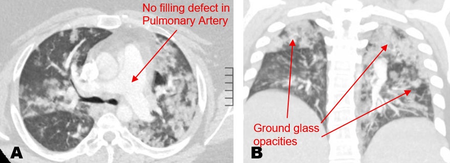
A 31-year-old woman with a history of antiphospholipid syndrome complicated by chronic pulmonary embolism on chronic warfarin therapy with target INR of 2–3, obstructive sleep apnea, primary sclerosing cholangitis on ursodiol, and body mass index (BMI) 58 presented to the emergency department with worsening dyspnea and nonproductive cough. She has had a history of recurrent pneumonia for the past two years; however, each one would be resolved with antibiotics. She was recently hospitalized two months prior for severe sepsis due to pneumonia and required intermittent use of 2 L of oxygen on discharge. However, she came in subsequently requiring 6 L of oxygen with shortness of breath. Her INR was 3, with this her warfarin was continued and titrated to meet her INR goal. She received a computed tomography angiography (CTA) of the chest that showed findings of multifocal pneumonia (Figure 1). Her infectious workup was completely negative. Inflammatory and sepsis related laboratory markers were not strongly suggestive with values showing ESR 61 mm/h, C-Reactive Protein (CRP) < 2.9 mg/dL, lactate 0.7 mmol/L, procalcitonin 0.7 ng/mL, and white blood cell count 5.3 × 10⁹/L, raising concern for alternative process.
Yet she completed a 7-day course of cefepime and doxycycline, but due to the severity of imaging and worsening respiratory status, cardiothoracic surgery was consulted for a video-assisted thoracoscopic surgery biopsy on the right side with wedge resection of the right upper lobe, as there was suspicion of an autoimmune process, to which she was subsequently started on Prednisone 40 mg daily. Lab work revealed positive ANA and P-ANCA titers. A few days later, the pathology report showed diffuse intra-alveolar hemosiderin-laden macrophages and interstitial hemosiderin deposition with neutrophilic infiltrate in the alveolar septa, consistent with capillaritis. From the pathology and history of antiphospholipid syndrome, this was the likely etiology of the patient’s lung hemorrhage. In addition, her myeloperoxidase and Proteinase 3 were negative. She was stable on the Prednisone 40 mg daily and discharged home with a stable respiratory status but had plans of weaning off from it with the introduction of Rituximab.
After getting her first dose of Rituximab 1000 mg in 0.9% NaCl 600 mL infusion, she developed further shortness of breath. This was assumed, possibly due to the taper of Prednisone and Rituximab failure or even infection. Thus, bronchoscopy was planned to rule out infection with the complexity of her condition as there was suspicion of Pneumocystitis carinii pneumonia (PJP), and even to identify if there is current DAH. Rheumatology was consulted and discussed that with CD20 check being less than 1, this was considered Rituximab failure as CD20 was appropriately depleted. Steroids were given intravenously (IV) with a Prednisone equivalent of 60 mg with IV Solumedrol 125 mg twice daily. She had a negative infectious workup, stabilized to her baseline, and had follow up visit at Mayo Clinic for further workup.
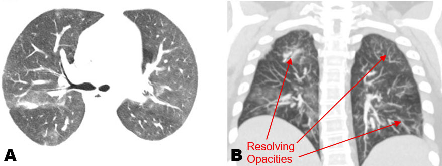
At Mayo Clinic, workup showed that her presentation was an uncommon presentation of anti-phospholipid syndrome, and the decision was made to start her on cyclophosphamide. She had an extended stay in the hospital with cyclophosphamide 1000 mg infusion but was only able to complete 2 cycles of treatment and received plasma exchange ×5 to remove potential circulating antiphospholipid antibodies and inflammatory mediators. After a prolonged stay, she was stable with markedly improved computed tomography (CT) chest (Figure 2). Shortly after, the patient was discharged to inpatient rehabilitation. She improved remarkably in the inpatient rehabilitation facility and was discharged back home. She continues to be on immunosuppressive therapy with Prednisone 30 mg twice a day, with plans for multidisciplinary outpatient follow-up.
Discussion
This case represents a rare but severe pulmonary manifestation of antiphospholipid syndrome of diffuse alveolar hemorrhage, thought to result from immune-mediated injury to the pulmonary vasculature [3]. The significance of this case shows the complexity in diagnosis and management of DAH associated with APS, especially with the high relapse rate of 52% within the first year [6]. This patient underwent frequent readmissions to the hospital, thought to be a result of pneumonia, especially as hemoptysis was absent in her disease course, leading to the delay in recognition of findings. However, presumed recurrent pneumonia with persistently negative infectious evaluation ultimately prompted surgical lung biopsy, which demonstrated pulmonary capillaritis with hemosiderin-laden macrophages consistent with DAH. This highlights the importance of considering DAH in patients with APS who present with recurrent pulmonary infiltrates and progressive hypoxemia despite appropriate antimicrobial therapy.
Despite biopsy-confirmed pulmonary capillaritis consistent with diffuse alveolar hemorrhage, the patient underwent further evaluation for alternative autoimmune etiologies. Laboratory testing revealed a positive p-ANCA; however, this was considered a false-positive result given negative myeloperoxidase antibodies and the absence of clinical features suggestive of ANCA-associated vasculitis [7]. False-positive p-ANCA results have been reported in association with primary sclerosing cholangitis and may reflect underlying immune dysregulation rather than true vasculitis [8]. Additional evaluation for systemic lupus erythematosus was unrevealing based on serologic testing and lack of supporting clinical features [9]. This case highlights the importance of maintaining a broad autoimmune differential when evaluating diffuse alveolar hemorrhage in patients with antiphospholipid syndrome, as accurate etiologic identification directly informs immunosuppressive treatment selection.
High-dose steroids remain first-line therapy for APS-associated DAH; however, relapse during steroid tapering is common and frequently necessitates a bridge to steroid-sparing immunosuppressive agents [3]. In this patient, when she was being tapered from her steroids, she had exacerbations in her condition and was started on Rituximab. This had treatment failure as her CD20 check was less than 1, showing that the CD20 was appropriately depleted [10]. From this, evaluation at a tertiary center helped to further guide management decisions, emphasizing the importance of multidisciplinary collaboration in complex APS-associated pulmonary disease. She was eventually started on treatment with Cyclophosphamide, to which her condition stabilized after two cycles during inpatient admission with plasma exchange to help remove circulating antiphospholipid antibodies and reduce immune complexes.
Favorable findings in this patient’s condition was the lack of high-risk APS phenotype denoted by persistently high Lupus Anticoagulant, Anticardiolipin antibodies, and Anti-β2 glycoprotein antibodies [11]. She also had no thrombocytopenia and no findings of catastrophic APS as she only had pulmonary involvement [12]. Patients with such features have been associated with increased relapse rates and higher long-term mortality in APS-associated diffuse alveolar hemorrhage despite immunosuppressive therapy [6]. Following escalation of immunosuppressive therapy, the patient’s respiratory status stabilized, and she was discharged on a tapering regimen of systemic corticosteroids with plans for continued outpatient multidisciplinary follow-up.
Conclusion
This case highlights the importance of maintaining clinical suspicion for APS-associated DAH in patients with persistent pulmonary infiltrates despite negative infectious evaluation. Early recognition is critical as diffuse alveolar hemorrhage is a rare but life-threatening pulmonary manifestation of antiphospholipid syndrome that may mimic infection and delay diagnosis. This case underscores the importance of early diagnostic evaluation for pulmonary capillaritis in patients with APS and persistent hypoxemic respiratory failure as treatment-refractory disease may require escalation to cyclophosphamide and adjunctive therapies such as plasma exchange.
LEARNING POINTS
- APS-associated DAH should be considered in patients with APS presenting with recurrent pulmonary infiltrates and unexplained hypoxemia despite negative infectious workup
- Refractory disease may require escalation beyond corticosteroids and rituximab to cyclophosphamide and adjunctive therapies such as plasma exchange
- Hemoptysis may be absent early in diffuse alveolar hemorrhage and bronchoscopy, or lung biopsy may be required to establish the diagnosis in patients with persistent radiographic abnormalities
- Early recognition of severe or treatment-refractory disease is critical, as APS-associated diffuse alveolar hemorrhage is associated with high relapse rates and substantial long-term morbidity despite immunosuppressive therapy
REFERENCES
1.
Rout P, Goyal A, Singhal M. Antiphospholipid syndrome. In: StatPearls. Treasure Island (FL): StatPearls Publishing; 2026.
[Pubmed]

2.
Stoots SA, Lief L, Erkan D. Clinical insights into diffuse alveolar hemorrhage in antiphospholipid syndrome. Curr Rheumatol Rep 2019;21(10):56. [CrossRef]
[Pubmed]
3.
Park JA. Treatment of diffuse alveolar hemorrhage: Controlling inflammation and obtaining rapid and effective hemostasis. Int J Mol Sci 2021;22(2):793. [CrossRef]
[Pubmed]
4.
Madu A, Siddiqui AH. Idiopathic pulmonary hemorrhage. In: StatPearls. Treasure Island (FL): StatPearls Publishing; 2026.
[Pubmed]
5.
Cartin-Ceba R, Peikert T, Ashrani A, Keogh K, Wylam ME, Ytterberg S, et al. Primary antiphospholipid syndrome-associated diffuse alveolar hemorrhage. Arthritis Care Res (Hoboken) 2014;66(2):301–10. [CrossRef]
[Pubmed]
6.
Figueroa-Parra G, Meade-Aguilar JA, Langenfeld HE, González-Treviño M, Hocaoglu M, Hanson AC, et al. Clinical features, risk factors, and outcomes of diffuse alveolar hemorrhage in antiphospholipid syndrome: A mixed-method approach combining a multicenter cohort with a systematic literature review. Clin Immunol 2023;256:109775. [CrossRef]
[Pubmed]
7.
Kitching AR, Anders HJ, Basu N, Brouwer E, Gordon J, Jayne DR, et al. ANCA-associated vasculitis. Nat Rev Dis Primers 2020;6(1):71. [CrossRef]
[Pubmed]
8.
Roozendaal C, Van Milligen de Wit AW, Haagsma EB, Horst G, Schwarze C, Peter HH, et al. Antineutrophil cytoplasmic antibodies in primary sclerosing cholangitis: Defined specificities may be associated with distinct clinical features. Am J Med 1998;105(5):393–9. [CrossRef]
[Pubmed]
9.
Al-Adhoubi NK, Bystrom J. Systemic lupus erythematosus and diffuse alveolar hemorrhage, etiology and novel treatment strategies. Lupus 2020;29(4):355–63. [CrossRef]
[Pubmed]
10.
Hartinger JM, Kratky V, Hruskova Z, Slanar O, Tesar V. Implications of rituximab pharmacokinetic and pharmacodynamic alterations in various immunemediated glomerulopathies and potential anti-CD20 therapy alternatives. Front Immunol 2022;13:1024068. [CrossRef]
[Pubmed]
11.
Schulman S, Sholzberg M, Spyropoulos AC, Zarychanski R, Resnick HE, Bradbury CA, et al. ISTH guidelines for antithrombotic treatment in COVID-19. J Thromb Haemost 2022;20(10):2214–25. [CrossRef]
[Pubmed]
12.
Bitsadze V, Yakubova F, Khizroeva J, Lazarchuk A, Salnikova P, Vorobev A, et al. Catastrophic antiphospholipid syndrome. Int J Mol Sci 2024;25(1):668. [CrossRef]
[Pubmed]
SUPPORTING INFORMATION
Author Contributions
Srujan Kancharla - Conception of the work, Design of the work, Acquisition of data, Analysis of data, Drafting the work, Revising the work critically for important intellectual content, Final approval of the version to be published, Agree to be accountable for all aspects of the work in ensuring that questions related to the accuracy or integrity of any part of the work are appropriately investigated and resolved.
Claire Heigl Maza - Acquisition of data, Analysis of data, Drafting the work, Revising the work critically for important intellectual content, Final approval of the version to be published, Agree to be accountable for all aspects of the work in ensuring that questions related to the accuracy or integrity of any part of the work are appropriately investigated and resolved.
Jadvinder Singh Goraya - Acquisition of data, Analysis of data, Drafting the work, Revising the work critically for important intellectual content, Final approval of the version to be published, Agree to be accountable for all aspects of the work in ensuring that questions related to the accuracy or integrity of any part of the work are appropriately investigated and resolved.
Guaranter of SubmissionThe corresponding author is the guarantor of submission.
Source of SupportNone
Consent StatementWritten informed consent was obtained from the patient for publication of this article.
Data AvailabilityAll relevant data are within the paper and its Supporting Information files.
Conflict of InterestAuthors declare no conflict of interest.
Copyright© 2026 Srujan Kancharla et al. This article is distributed under the terms of Creative Commons Attribution License which permits unrestricted use, distribution and reproduction in any medium provided the original author(s) and original publisher are properly credited. Please see the copyright policy on the journal website for more information.


