
 |
Case Report
First reported capitellum fracture in a rare combination of Galeazzi fracture with elbow dislocation: A case report and literature review
1 Department of Orthopaedic Surgery, Aalborg University Hospital, Aalborg, Denmark
2 Shoulder Section, Department of Orthopaedic Surgery, Aalborg University Hospital, Farsoe, Denmark
Address correspondence to:
Hasan Zalzala
Department of Orthopaedic Surgery, Aalborg University Hospital, Aalborg,
Denmark
Message to Corresponding Author
Article ID: 100033Z14HZ2026
Access full text article on other devices

Access PDF of article on other devices


How to cite this article
Zalzala H, Alrubaie R, Jensen SL. First reported capitellum fracture in a rare combination of Galeazzi fracture with elbow dislocation: A case report and literature review. J Case Rep Images Orthop Rheum 2026;8(2):1–5.ABSTRACT
Introduction: Galeazzi fractures, defined as a radial shaft fracture with distal radioulnar joint (DRUJ) dislocation, are uncommon injuries in adults. Their combination with ipsilateral elbow dislocation is rare, with only a limited number of cases reported. We present the first documented case of a Galeazzi fracture with elbow dislocation combined with a capitellum fracture, alongside additional associated injuries.
Case Report: A 35-year-old male sustained a high energy trauma following a fell from the second floor (approximately 8 meters). Clinical examination revealed pain and deformity of the left elbow and wrist without neurovascular compromise. Imaging demonstrated a Galeazzi fracture, elbow dislocation, scaphoid fracture, and, following reduction, a capitellum fracture with an associated radial head fracture. Associated injuries included a T7 vertebral fracture and a grade II splenic laceration. The patient remained hemodynamically stable; therefore there was no indication for fluid or blood resuscitation. Initial management consisted of closed reduction of the elbow dislocation, followed by staged surgical intervention. The radial fracture was treated with open reduction and internal fixation (ORIF) and distal radioulnar joint (DRUJ) stabilization. A subsequent procedure addressed fixation of the scaphoid and capitellum fractures, along with repair of the lateral collateral ligament. At one-year follow-up, the patient demonstrated full elbow range of motion (ROM) without functional impairment. A residual limitation in forearm supination persisted and necessitation subsequent removal of the radial plate after one year.
Conclusion: This case describes a previously unreported combination of capitellum fracture, Galeazzi fracture, and elbow dislocation. Such injury patterns are typically associated with high-energy mechanisms, and awareness of concomitant injuries (vertebral fracture and spleen laceration in our case) is essential to avoid missed diagnoses. Despite the inherent complexity, staged surgical management can yield satisfactory functional outcomes.
Keywords: Capitellum fracture, Elbow dislocation, Galeazzi fracture
Introduction
Galeazzi fracture is defined as a fracture of the radius with dislocation of the distal radioulnar joint (DRUJ). It accounts for approximately 3–7% of all forearm fractures in adults [1]. The elbow dislocation rate is 6–13 cases per 100,000 people and accounts for 11–28% of all injuries to the elbow [2]. The combination of a Galeazzi fracture with elbow dislocation in the same extremity is rarely reported, with only 10 reported cases in the literature [3],[4],[5],[6],[7],[8],[9],[10],[11],[12]. We report a unique case of a Galeazzi fracture associated with posterolateral dislocation of the elbow that is combined with several other injuries, including a capitellum fracture, representing the first documented occurrence of this specific fracture configuration and injury pattern.
Case Report
A 35-year-old male fell approximately 8 meters from his apartment window on the first floor. He was under the influence of alcohol, rendering him unable to recall the exact trauma mechanism.
Clinical Findings
Past medical history and comorbidities
The patient had no known chronic medical conditions, allergies, or previous surgical history relevant to the current presentation.
Upon examination, the patient reported pain in his left elbow and wrist. Physical examination revealed swelling with ecchymosis on the anterior and lateral aspects of the elbow. The arm was held in mild flexion with a visible deformity characterized by loss of the natural olecranon prominence and a clear triangular deformity evident when viewed from the lateral aspect. No visible wounds were noted on inspection of the upper extremity. Neurovascular examination was intact with no deficits noted.
Vital signs and initial hemodynamic status
The patient presented with stable vital signs: blood pressure 137/89 mmHg, heart rate 98 beats per minute, temperature 37.2°C, and respiratory rate 15 breaths per minute. No fluid resuscitation or blood transfusion was required during the acute management phase.
Diagnostic Assessment
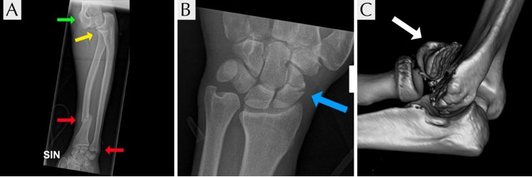
Initial plain radiographs of the left upper extremity demonstrated a radial shaft fracture with associated elbow and DRUJ dislocations, as well as a scaphoid fracture (Figure 1). Following closed reduction of the elbow, computed tomography (CT) scan was obtained to further reveal the displaced capitellum fracture and undisplaced radial head fracture. Additionally, a trauma CT scan identified a T7 vertebral fracture and grade II splenic injury according to the American Association for the Surgery of Trauma (AAST) splenic injury scale without contrast extravasation (Figure 2).
Therapeutic intervention
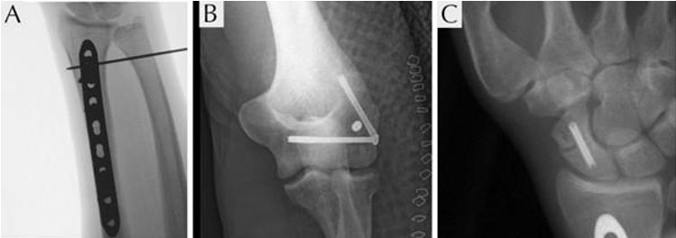
Closed reduction of the elbow was performed after 4 hours under general anesthesia. Surgical intervention on the Galeazzi fracture was performed one day after the injury with open reduction and internal fixation (ORIF) of the radius using a 3.5-millimeter Locking Compression Plate (LCP) through a modified Henry approach. Moreover, transfixation of the distal radioulnar joint (DRUJ) with 1.6 mm K-wire was performed (Figure 3A). Surgery for the scaphoid and capitellum fractures was performed eight days after the injury when specialist service was available. Perioperatively, after removing the transfixation K-wire, the DRUJ was tested and found stable, with no further need for transfixation. The capitellum fracture was osteosynthesized using a 3.5 mm cannulated screw supplemented with two headless compression screws via a Kaplan approach. The lateral collateral ligament, which was found disrupted, was reinserted with an all-suture anchor on the lateral humeral epicondyle (Figure 3B). The fracture of the radial head was left untouched. The elbow was visualized under fluoroscopy and found stable with a congruent joint. The scaphoid was osteosynthesized with a headless compression screw via a minimally invasive palmar approach (Figure 3C).
The splenic injury was managed conservatively with observation, as no contrast extravasation or hemodynamic instability was present. No blood transfusion or surgical intervention was required.
Postoperative care
The extremity was immobilized for three weeks in a long arm cast at 90° elbow flexion and thereafter active, unloaded movement was allowed in a stabilizing hinged brace.
Follow-up and outcomes
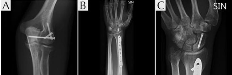
At follow-up after eight weeks, the patient reported no pain. The elbow range of motion (ROM) was measured 5–130°, supination 0–45°, and pronation 0–60°. Full ROM was observed in the wrist. By 12 weeks, the patient maintained a limitation in supination (0–45°) but demonstrated improvement in pronation (0–60°). Physiotherapy was initiated after eight weeks, allowing full movement and weight-bearing. Final radiographs demonstrated stable fixation and healed fractures (Figure 4). At 1-year follow-up, the patient exhibited unrestricted elbow range of motion. Residual limitation of wrist motion persisted, leading to subsequent removal of the LCP on the radius.

Discussion
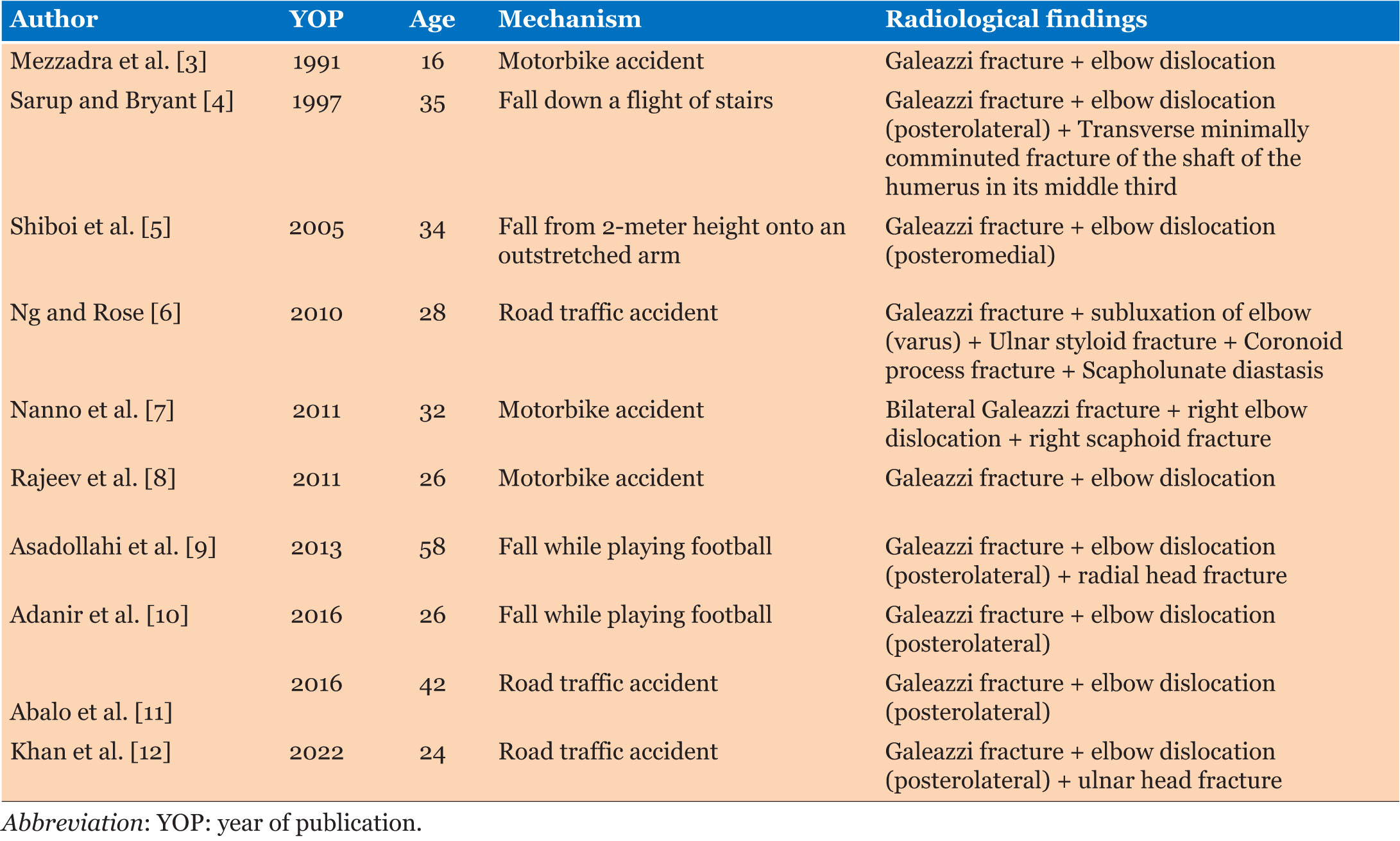
A literature review by Ng and Rose [6] and Asadollahi et al. [9] have previously documented cases of Galeazzi fractures occurring in combination with elbow dislocation. For our review, we performed a comprehensive search of the PubMed, Embase, and Scopus databases using the search term (“Galeazzi fracture” AND (“Elbow dislocation” OR “Elbow joint dislocation”)). We also conducted a manual citation search to ensure the inclusion of all relevant publications. Including the cases presented in the two reviews, our search yielded a total of 10 reported cases, 5 of which featured concurrent ipsilateral fractures around the elbow or wrist (Table 1).
Our case describes a similar injury pattern but is unique due to the presence of three associated injuries (scaphoid fracture, radial head fracture, and a capitellum fracture). Notably, capitellum fracture in association with this injury pattern has not been previously reported in the literature.
In previously reported cases, this type of injury is commonly associated with high-energy trauma, such as falls from significant heights or road traffic accidents (Table 1). Although the patient’s alcohol intoxication hindered us from determining the precise mechanism of the elbow injury, we hypothesize that it mirrors the mechanism described by Asadollahi et al. [9] and Sarup and Bryant [4]. This mechanism involves extreme axial loading on an outstretched, pronated hand, driven by the patient’s body weight during the fall.
Awareness of potentially associated injuries in high-energy trauma patients is crucial, as missed injuries can result in delayed treatment and poorer clinical outcomes. Treatment of Galeazzi fracture, elbow dislocation, or other associated injuries does not change despite their concurrent occurrence, as every injury is managed on its own terms [5],[11]. Anatomic ORIF of the radius is the standard treatment for most patients with Galeazzi fracture [1]. Further intraoperative interventions such as transfixation are based on the reducibility and post-reduction stability of the DRUJ [5]. Elbow dislocation is treated with closed reduction, but immediate open reduction of the elbow is indicated in the cases of complex elbow fracture-dislocation where closed reduction has failed.
In our review of the literature, we found that all cases with Galeazzi fracture were treated successfully with ORIF. Elbow dislocation was treated with closed reduction in all except 2 cases (by Rajeev et al. [8] and Ng and Rose [6]), where open reduction was indicated. The postoperative follow-up period in the literature ranges from five months to seven years. Across all cases, elbow range of motion was preserved within acceptable limits without functional impairment. However, our patient demonstrated a residual limitation in forearm supination, leading to subsequent removal of the LCP from the radius.
Conclusion
This case represents the first documented occurrence of a capitellum fracture in association with a Galeazzi fracture and ipsilateral elbow dislocation. This complex injury pattern highlights the critical importance of comprehensive imaging and systematic evaluation in high-energy trauma patients to identify all associated injuries and prevent missed diagnoses. Although the patient presented with multiple severe injuries involving the upper extremity, vertebral column, and solid organs, appropriate staged surgical management with attention to fracture reduction, soft tissue repair (lateral collateral ligament), and DRUJ stabilization resulted in satisfactory functional recovery. Residual limitation in forearm supination led to eventual plate removal, emphasizing the need for careful postoperative assessment and willingness to modify treatment based on functional outcomes. Awareness of this rare injury pattern and similar polytrauma presentations will enable orthopedic surgeons to optimize patient outcomes through early recognition and appropriate surgical planning.
REFERENCES
1.
Atesok KI, Jupiter JB, Weiss APC. Galeazzi fracture. J Am Acad Orthop Surg 2011;19(10):623–33. [CrossRef]
[Pubmed]

2.
Hobgood ER, Khan SO, Field LD. Acute dislocations of the adult elbow. Hand Clin 2008;24(1):1–7. [CrossRef]
[Pubmed]
3.
Mezzadra A, Gusmeroli E, Tettamanzi M, Prestianni B, Molinari D. A rare injury to the upper limb: Elbow dislocation combined with Galeazzi fracture-dislocation. Ital J Orthop Traumatol 1991;17(4):567–72.
[Pubmed]
4.
Sarup S, Bryant PA. Ipsilateral humeral shaft and Galeazzi fractures with a posterolateral dislocation of the elbow: A variant of the “floating dislocated elbow.” J Trauma 1997;43(2):349–52. [CrossRef]
[Pubmed]
5.
Shiboi R, Kobayashi M, Watanabe Y, Matsushita T. Elbow dislocation combined with ipsilateral Galeazzi fracture. J Orthop Sci 2005;10(5):540–2. [CrossRef]
[Pubmed]
6.
Ng VY, Rose S. Combined elbow fracture-dislocation and Galeazzi fracture in an adult: A case report and review of the literature. Current Orthopaedic Practice 2010;21(3):330–3. [CrossRef]
7.
Nanno M, Sawaizumi T, Takai S. Case of bilateral Galeazzi fractures associated with dislocation of the right elbow. J Nippon Med Sch 2011;78(6):384–7. [CrossRef]
[Pubmed]
8.
Rajeev A, Senevirathna S, Harrison J. Subacute dislocation of the elbow following Galeazzi fracture-dislocation of the radius: A case report. J Med Case Rep 2011;5:589. [CrossRef]
[Pubmed]
9.
Asadollahi S, Shepherd D, Hau RC. Elbow fracture-dislocation combined with Galeazzi fracture in adult: A case report and literature review. Int J Surg Case Rep 2013;4(8):748–52. [CrossRef]
[Pubmed]
10.
Adan?r O, Yüksel S, Beytemur O, Güleç MA. Open Galeazzi fracture with ipsilateral elbow dislocation. Eklem Hastalik Cerrahisi 2016;27(2):113–6. [CrossRef]
[Pubmed]
11.
Abalo A, Akloa KK, Dellanh YY. Elbow dislocation with ipsilateral Galeazzi fracture: A case report. Open Journal of Orthopedics 2016;6(9):276–82. [CrossRef]
12.
Khan MJ, Raza A, Singhal U, Asif S. Combination of Galeazzi fracture, fracture ulnar head, and ipsilateral posterior elbow dislocation: A rare case report. J Bone Joint Dis 2022;37(1):15–7. [CrossRef]
SUPPORTING INFORMATION
Acknowledgments
Artificial Intelligence (AI) Disclosure
Artificial intelligence language models (Claude and ChatGPT) were used in this manuscript to assist with grammar checking, spelling correction, and text formatting to improve clarity and presentation. However, all clinical data, patient information, case observations, medical analysis, and scientific content are original work of the authors. The AI tools were not used for clinical decision-making, data interpretation, or generation of medical content.
Hasan Zalzala - Conception of the work, Design of the work, Acquisition of data, Drafting the work, Revising the work critically for important intellectual content, Final approval of the version to be published, Agree to be accountable for all aspects of the work in ensuring that questions related to the accuracy or integrity of any part of the work are appropriately investigated and resolved.
Reem Alrubaie - Conception of the work, Design of the work, Acquisition of data, Drafting the work, Revising the work critically for important intellectual content, Final approval of the version to be published, Agree to be accountable for all aspects of the work in ensuring that questions related to the accuracy or integrity of any part of the work are appropriately investigated and resolved.
Steen Lund Jensen - Conception of the work, Design of the work, Acquisition of data, Drafting the work, Revising the work critically for important intellectual content, Final approval of the version to be published, Agree to be accountable for all aspects of the work in ensuring that questions related to the accuracy or integrity of any part of the work are appropriately investigated and resolved.
Guaranter of SubmissionThe corresponding author is the guarantor of submission.
Source of SupportNone
Consent StatementWritten informed consent was obtained from the patient for publication of this article.
Data AvailabilityAll relevant data are within the paper and its Supporting Information files.
Conflict of InterestAuthors declare no conflict of interest.
Copyright© 2026 Hasan Zalzala et al. This article is distributed under the terms of Creative Commons Attribution License which permits unrestricted use, distribution and reproduction in any medium provided the original author(s) and original publisher are properly credited. Please see the copyright policy on the journal website for more information.


